
FIGURE 2.
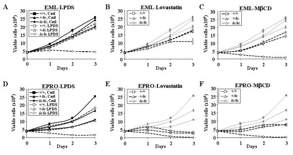
Loss of Lbr expression in EML and EPRO-+/ic and –ic/ic cells suppresses growth responses in regular medium or under cholesterol deprivation conditions. EML cells were plated at 2 × 10⁵ cells/ml in 2 ml medium that contained (A) normal serum (Ctrl) or lipoprotein deficient serum (LPDS), (B) normal serum plus 10μM lovastatin, or (C) normal serum plus 5mM Methyl β-Cyclodextrin (MβCD), and daily counts of viable cells were performed. Data shown are from multiple assays performed simultaneously, in which cells were plated in 2 ml of medium with each condition using triplicate plates per assay; the data from cells grown in regular medium are displayed in (A) as black lines and then shown as grey lines in (B) and (C) to highlight the changes of growth in cholesterol depleted conditions. Similar studies were performed with EPRO cells derived from the EML cells of each genotype, using (D) normal serum vs. LPDS, (E) lovastatin or (F) MβCD. Viable cells were identified by trypan blue exclusion and increases in total cell numbers was determined manually using a hemacytometer at 24-hour intervals for three days. Data represent results from at least two independent assays.