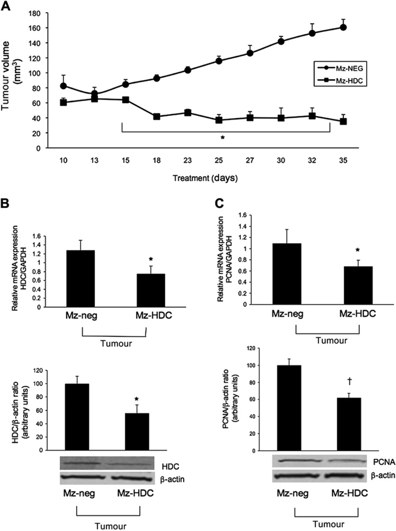
Figure 7.
(A) Xenograft tumour growth over time after implantation of genetically modified Mz-ChA-1 cells. After 18 days the Mz-HDC tumours decreased in volume and remained similar throughout the measurement time compared with the Mz-neg tumours which continued steadily to increase in volume. By two-way ANOVA, tumour growth in Mz-HDC was significantly lower (p<0.001) than in Mz-neg at all time points except day 13. (B,C) By real-time PCR and immunoblots in RNA and protein samples from tumours extracted from both Mz-neg and Mz-HDC, a significant decrease was found in histidine decarboxylase (HDC) expression in Mz-HDC tumour cells compared with Mz-neg tumour cells and proliferating cellular nuclear antigen (PCNA) expression (*p<0.05 vs Mz-neg cells; †p<0.01 vs Mz-neg cells). Data are mean±SEM of three experiments (real-time PCR) and eight experiments (immunoblotting).

