
Figure 1. VV infection in mast cell-deficient Kit wsh−/− mice.
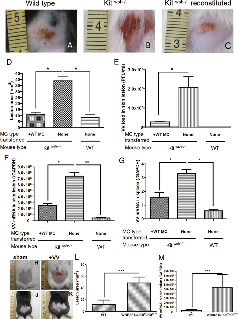
106 PFU of VV was applied on the lower backs of Kitwsh−/− mice and their wild-type littermate control mice. (A) Left image is representative of the reaction seen at 72 hours in wild type mice. (B) The center image is representative of the skin lesion in Kitwsh−/− mice. (C) The right image is Kitwsh−/− mice reconstituted with mast cells. (D) Quantification of lesion size (mm2) at 72 h post-infection. (E) Quantification of viral load (in PFU/ml) in the skin lesions at 72 h post-infection. (F) Expression of VV early gene at the skin lesions was quantified by real-time quantitative RT-PCR. (G) Expression of VV early gene in the spleen was quantified by real-time quantitative RT-PCR. *P<0.05. VV infection in mast cell-deficient WBB6F1/J-KitW/KitW-v double heterozygotes mice (H–M) 106 PFU of VV was applied on the lower backs of WBB6F1/J-KitW/KitW-v double heterozygotes mice and their wild-type littermate control mice. (H) Sham control of wild type mice scarified without VV application at 72 hours (I) Upper right image is representative of the skin lesion in WBB6F1/J-KitW/KitW-v double heterozygotes mice. (J) Sham control of WBB6F1/J-KitW/KitW-v double heterozygotes mice scarified without VV application at 72 hours (K) Lower right image is representative of the reaction seen at 72 hours in wild type mice. The right image is Kitwsh−/− mice reconstituted with mast cells. (L) Quantification of lesion size (mm2) at 72 h post-infection. (M) Expression of VV early gene at the skin lesions was quantified by real-time quantitative RT-PCR at 72 h post-infection.
Three independent experiments with 5 mice per group were performed for each experiment