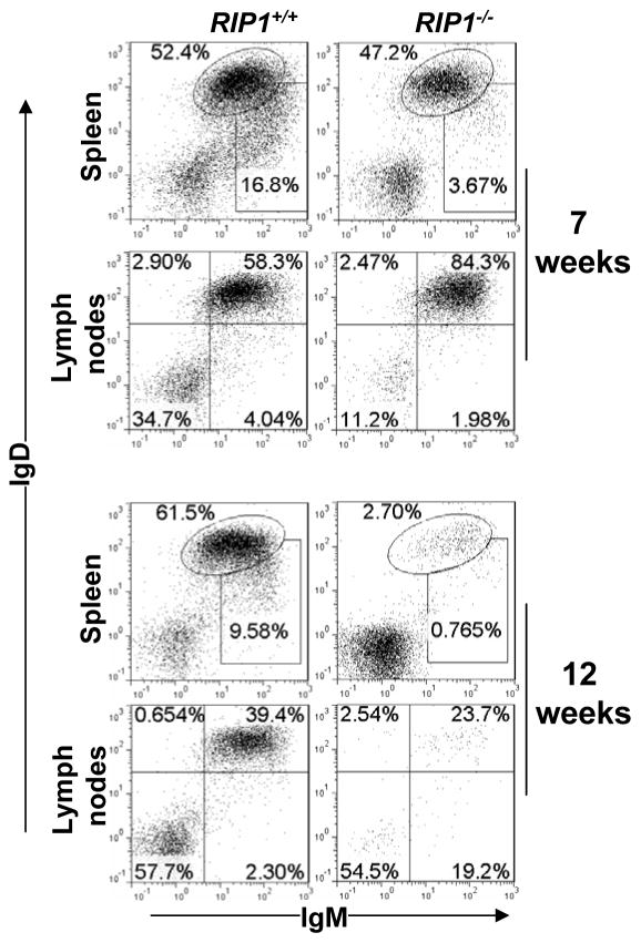
Fig. 2. RIP1 deficiency results in a time-dependent decline in the size of the peripheral B cell pool.
RIP1−/− fetal liver cells were adoptively transferred to immunodeficient NSG recipient mice irradiated (200 RAD), as described previously (25, 77). At 7 or 12 weeks post transfer, the resulting hematopoietic chimeras were used to prepare single cell suspensions from the specified lymphoid organs. Reconstitution of the peripheral B cell compartment was determined by flow cytometric analysis of the IgM+ and/or IgD+ populations in the indicated organs. Gates were set to indicate immature (IgDloIgM+) and mature (IgD+IgM+) B cell populations. The numbers are percentages of the gated populations. Data are representative of 5 independent experiments. RIP1+/+ fetal liver cell-transferred recipients were used as a control.

