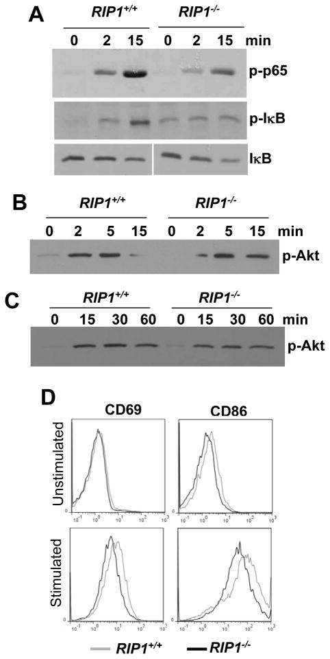
Fig. 5. LPS-induced signaling in RIP1−/− B cells.
Peripheral B cells purified from NSG recipients transferred with RIP1−/− or RIP1+/+ fetal liver cells (A and B) or MEFs of the indicated genotypes (C) were stimulated with LPS (10 μg/ml), and western blotting analysis was performed using Abs specific for p-p65 NF-κB, p-IκB, IκB, and p-Akt. D, The CD69 and CD86 activation markers on B cells were analyzed by flow cytometry 12 hours post stimulation with LPS (1 μg/ml). Unstimulated B cells were used as a control. Data shown are representative of at least 4 independent experiments.

