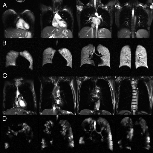
Figure 2:
Selected sections from representative 129Xe ventilation and 1H MR anatomic images in individual subjects. A, Steady-state free-precession 1H MR images in a healthy volunteer. B, Corresponding 129Xe ventilation MR images in the same healthy volunteer. C, Steady-state free-precession 1H MR images in a subject with COPD. D, Corresponding 129Xe ventilation MR images in the same subject with COPD show substantial ventilation defects and regions lacking ventilation.

