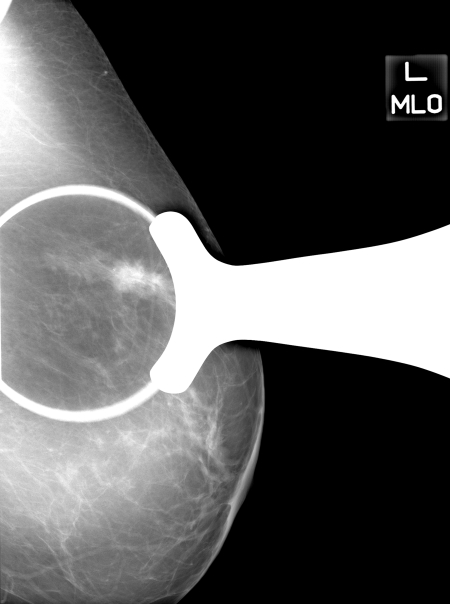
Figure 1c:
Images in 71-year-old woman with 1.9-cm invasive ductal carcinoma. (a) Cropped mediolateral oblique DBT image (arrow) displayed on the graphical user interface, which allowed readers to electronically enter their assessment of mass visibility, likelihood of malignancy, and BI-RADS category. This interface facilitated DBT image review by allowing readers to scroll through the image volume and adjust contrast and brightness as necessary. (b) MSV available on the graphical user interface to allow readers to electronically mark or reference the mass and (c) digital mediolateral oblique MSV (spot compression) reviewed by using a light-box or hot-light.

