
Figure 4b:
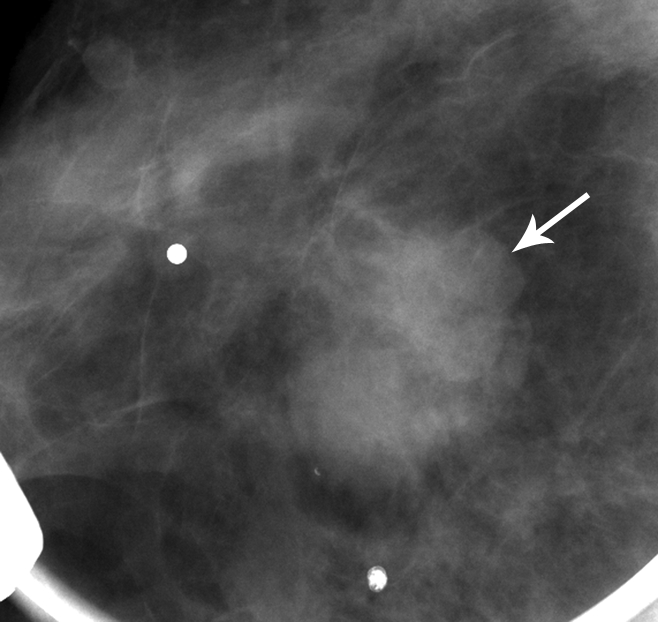
Matched image set of 1.2-cm fibroadenoma (arrow) in 29-year-old woman. (a) Mediolateral oblique DBT image (1-mm section from a volume of 20–30 sections) and (b) digital mediolateral oblique MSV (spot compression). The images were viewed and assessed during separate reader sessions. The readers’ mean and median visibility ratings, respectively, were 4.8 and 5.0 on DBT image and 5.5 and 4.5 on MSV (1 = obvious, 10 = subtle).