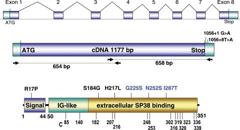
Figure 1.
ZPBP1 gene and cDNA and point mutations identified in teratozoospermic patients. ZPBP1 gene, cDNA amplification scheme and mutations in the 3′UTR are shown in the upper two diagrams. The position of the missense mutations in the ZPBP1 protein is shown in the lower diagram. The predicted protein structure is based on Interproscan, Prosite, Pfam and Blocks algorithms. ZPBP1 has a signal peptide (aa 1–44) and an extracellular region consisting of two predicted overlapping domains, an immunoglobulin-like (IG; aa 50–156) domain and an sp38 domain (aa 86–351). Homozygous mutations are depicted in blue, and heterozygous mutations are depicted in black. The positions of the cysteines (C) are shown at the bottom.

