Fig. 3.
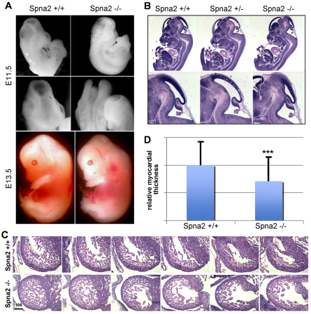
Spna2−/− embryos die at E12.5–16.5 with multiple defects. (A,B) Whole mounts depicting the gross morphological defects in head development, with frequent neural tube closure defects (E11.5 embryos shown). In cases where the neural tube closed, there remained distinct alterations in head and back curvature with craniofacial abnormalities. (C) Serial sections through the fetal heart. The Spna2−/− hearts have an irregular shaped ventricle, and a thinned compact myocardium. Scale bar: 100 μm. (D) Quantitative comparison of cardiac wall thickness. On average, the myocardium of the Spna2−/− hearts was 70.9% of the thickness of normal hearts of the same gestational age. This difference is highly significant (n=101; ***P=4.2×10⁻¹³). Error bars indicate s.d.

