Fig. 4.
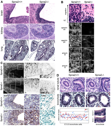
Histology of Spna2−/− embryos. (A) The VZ and SVZ of the developing brain display significant histological changes. Wild-type embryos at this stage of development display a thick pseudostratified VZ with active upward migration and population of overlying layers from the SVZ. In the Spna2−/− animals, the VZ was more compact, with fewer cells filling the more superficial layers (boxed area, shown enlarged in B). Other organs revealed no significant histological changes; kidney and lung are shown. In these organs, tubules and bronchioles were developing normally. (B) Enlargement of area outlined in A. In wild-type animals, αII- and βII-spectrin are rich in axon bundles (boxed) and are also present at synaptic terminals and initial axon segments (arrows). Ankyrin B is associated with unmyelinated axonal processes in the embryonic mouse (Chan et al., 1993), whereas ankyrin G is found along axons and in dendrites and concentrates in the initial axon segments (Kordeli et al., 1995). The loss of αII-spectrin disrupts the localization of these proteins, especially along the axon bundles, which no longer are marked by either spectrin or ankyrin. (C) Top two rows: Staining of sections with anti-tau demonstrates developing axon bundles in the VZ. Magnified images (3×) of the boxed areas are shown on the right with inverse contrast. Note the foreshortened, sparse and more disordered axon segments and increased concentration of tau near the soma when αII-spectrin is disrupted. Bottom two rows: Immunostaining for Ki67, a proliferation marker, reveals increased proliferation with spectrin loss. Averaged over multiple fields, and scoring any detected Ki67 staining as positive, 34±11% of cells in the wild-type animals were positive as compared with 46±8% of the cells in the spectrin-deleted animals. This represents a highly significant (P<0.006) 35% increase in proliferative activity. TUNEL staining (right) reveals no change in the level of apoptosis. (D) Higher power views of developing renal tubules and lung bronchioles indicate preservation of epithelial morphology. Comparison of epithelial cell height reveals no changes, even without laterally associated spectrin or ankyrin (see Fig. 5).

