Fig. 5.
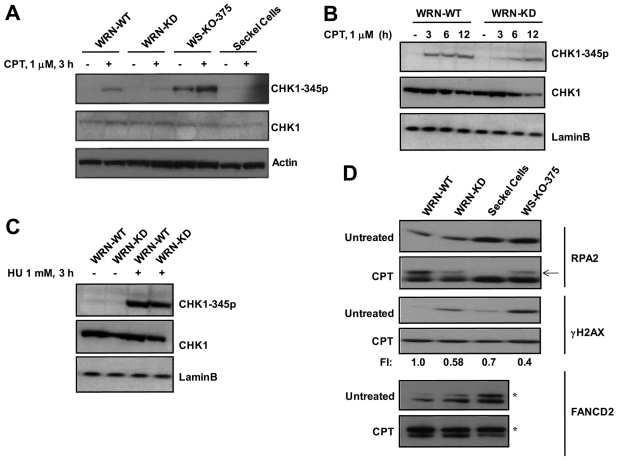
WRN regulates ATR-mediated signaling in response to CPT. (A) CHK1 phosphorylation on Ser345 was detected in WRN-WT, WRN-KD, WS-KO-375 cells and Seckel cells treated with CPT (1 μM). Cells were harvested at the indicated time point and cell lysates prepared as described for western analyses. (B) CHK1 phosphorylation was determined at various time points as indicated. (C) CHK1 phosphorylation on Ser345 was detected in control and WRN-KD cells treated with HU (1 mM, 3 hours). (D) Phosphorylation and ubiquitylation of ATR substrates in control and WRN-KD cells after CPT treatment (1 μM, 3 hours). Arrow shows phosphorylated form of RPA32; asterisk indicates monoubiquitylated form of FANCD2. Because endogenous levels of γH2AX and monoubiquitylated FANCD2 are high in WRN-depleted cells at untreated conditions, fold increase (FI) in the levels of induced γH2AX and monoubiquitylated FANCD2 in response to CPT are given by normalizing the fold increase (FI) in WRN-WT cells to 1.

