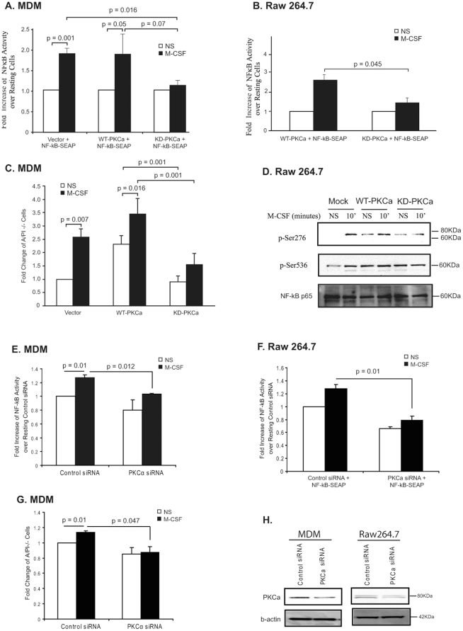
Figure 7. PKCα regulates phosphorylation of NF-κB p65 at Ser276.
(A) MDM or (B) RAW 264.7 cells were transiently transfected with pNF-κB-SEAP along with either WT-PKCα or the kinase-deficient (KD)-PKCα construct at a 1∶5 ratio. Cells were serum starved and stimulated with M-CSF and then SEAP secretion in the medium was measured. Data is from of three independent experiments. The p-value of cells transfected with KD compared to those transfected with WT was 0.05. (C) MDMs were removed from the plate using accutase and apoptosis of MDMs was measured by flow cytometry using Annexin V-FITC and propidium iodine (PI). (D) Whole cell lysates from the transfected RAW 264.7 cells were subjected to Western blot analysis with phospho-Ser276 or phospho-Ser536 NF-κB p65 antibodies. Blots were immunoblotted with PKCα to determine equal protein expression for the PKCα constructs. β-actin served as a loading control. Shown is a representative blot from three independent experiments. (E) MDM or (F) RAW 264.7 cells were transiently transfected with a pNF-κB-SEAP along with either 100 nM PKCα siRNA or control siRNA for 20-24 hours. Cells were serum starved for 2-4 hours and stimulated with 100 ng/ml M-CSF for 6 hours for MDM or RAW 264.7 for 2 hours and then SEAP secretion in the medium was measured. Shown is data of three independent experiments. (G) MDMs were removed from the plate using accutase and apoptosis of MDMs was measured by Annexin V-FITC and propidium iodine (PI) staining and analyzed by flow cytometry. (H) Whole cell lysates from the transfected MDM and RAW 264.7 cells were subjected to Western blot analysis with PKCα antibody. β-actin served as a loading control. Shown is a representative blot from at least three independent experiments.

