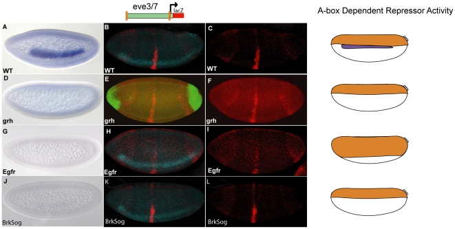
Figure 6. Analysis of A-box dependent and A-box independent repression in different mutant backgrounds.
Embryos (stage 5) were analyzed by in situ hybridiation for ind expression. Multiplex in situ hybridization was used to analyze A-box dependent and A-box independent repression in different mutant backgrounds. The schematic shows the CRMs used to drive expression of lacZ. The orange boxes in the schematic correspond to A-box sites while the orange boxes with a slash through them correspond to mutant A-box sites. The cartoons to the right of the images show where A-box/Cic dependent (orange) and A-box independent (green) repression are located in WT embryos and in the corresponding mutants; ind is only expressed in wildtype (purple). ind expression is shown in WT embryos (A), grh glc derived embryos (D), egfr mutants (G), and brk sog double mutants (J). The A-box-eve.stripe3/7-A-box reporter construct was introduced into different mutant backgrounds and analyzed by in situ hybridization; lacZ (red), and vnd (blue) is shown in a in WT embryo (B), grh glc derived embryo shows expression of hkb (green) rather than vnd and is tilted dorsally relative to the rest of the embryos (E), egfr mutant (H) and brk sog mutant (K). For clarity lacZ expression is shown alone for the corresponding embryos WT (C), grh glc (F), egfr mutant (I), and brk sog mutant (L). The same microscope settings were used to image C, I, and L; different settings were used for F but it was compared to a WT embryo taken under the same settings (not shown).

