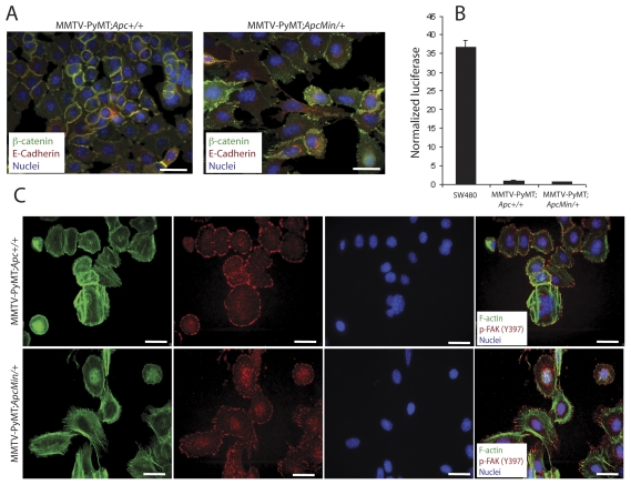
Figure 4. Cell lines generated from tumors have similar phenotypes to primary tumors.
Primary cells were isolated from mammary tumors from MMTV-PyMT;Apc+/+ and MMTV-PyMT;ApcMin/+ mice. (A) IF with a β-catenin antibody (green) and E-cadherin (red) antibody shows restricted localization of both proteins to cell-cell contacts in the control cells (left panel). In the Apc-mutant cells (right panel), junctional β-catenin and E-cadherin are observed but β-catenin is also localized in a punctate pattern in the cytosol or membrane. No nuclear accumulation of β-catenin is observed. Nuclei are stained with Hoechst (blue). Scale bars = 20 µm. (B) β-catenin/TCF reporter assays showed minimal basal Wnt/β-catenin pathway activation in both cell lines. SW480 cells were used as a positive control. The data are shown as a ratio of normalized TOPflash∶FOPflash values. (C) IF staining with phalloidin to label F-actin (green) illustrates a more spread morphology with extensive lamellipodia formation for the MMTV-PyMT;ApcMin/+ tumor cells (bottom panels) compared to the control cells (top panels). In addition, phospho-FAK (Tyr397, red) localizes in plaques primarily at the lamellipodia along actin stress fibers. Nuclei are stained with Hoechst (blue). Scale bars = 20 µm.

