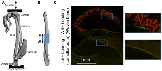
Figure 1. Mechanical loading was applied to the rat forelimb and a central region of the ulna was analyzed.
(A) Medial view of bones in a right forelimb of a rat obtained by microCT during simulated loading (Reprinted from Journal of Biomechanics, 40, Uthgenannt BA & Silva MJ, 317–324, 2007, with permission from Elsevier). (B) The central 5 mm of the ulna and surrounding periosteum were isolated for microarray analysis. (C) Representative transverse histological sections from a previous study [5] that illustrate bone formation after loading. WBF loading leads to woven bone formation while LBF loading increases lamellar bone formation. After loading, fluorochrome labels were injected in vivo on days 3 (green) and 8 (red) prior to animal sacrifice on day 10. Plastic embedded transverse sections were taken 1 mm distal to the ulna midpoint.

