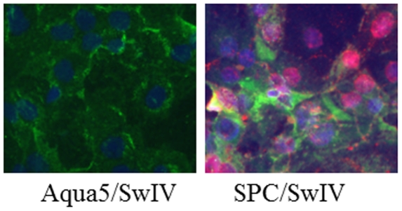
Figure 4. Replication of influenza virus in differentiated pneumocytes:
Primary cells were subcultured onto collagen I-coated plates in epithelial cell differentiation medium; on day 5 differentiated cells were infected with SwIV and examined for the presence of viral proteins at 24 h after infection. Type II pneumocytes, positive for SPC marker (green) supported the replication of SwIV virus as indicated by expression of viral NP protein (red). Cell nuclei were stained with DAPI (blue). No viral proteins were detected in Aqua5 expressing (green) type I pneumocytes.

