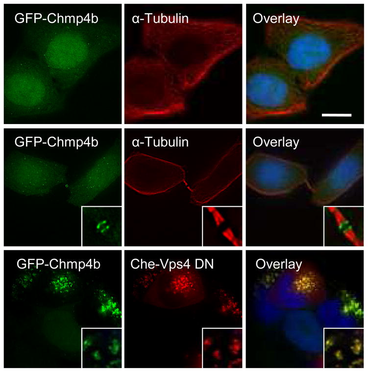
Figure 1. Characterization of the GFP-Chmp4b-expressing cell clone.
Hela cells stably expressing GFP-Chmp4b (green) were fixed and stained with anti-α-tubulin antibodies (red) and with DAPI (blue). Images show the distribution of GFP-Chmp4b in interphase cells (top panels) and in telophase cells (middle panels). Alternatively, cells were transfected with mCherry-Vps4-DN (bottom panels), fixed 18 hours post-transfection and stained with DAPI (blue). Samples were observed with an epifluorescence microscope. Deconvolved optical sections acquired at the center of the vertical dimension of the cell are shown. Expanded views are shown in insets. The scale bars represent 10μm.

