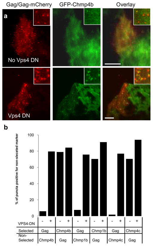
Figure 3. Catalytically inactive Vps4A increases localization of stably expressed GFP tagged ESCRT-III proteins at sites of HIV-1 assembly.
a. Hela cells stably expressing GFP-Chmp4b (green) were transfected with HIV-1 Gag/Gag-mCherry (red), in the absence (top panel) or presence (bottom panel) of Vps4A- DN. Cells were fixed 24 hours later and observed with a TIR-FM microscope. Expanded views are shown in insets. The scale bar represents 10μm.
b. Quantification of the co-localization between VLPs and puncta of ESCRT proteins. Hela cells stably expressing GFP-fused ESCRT-III proteins were transfected with Gag/Gag-mCherry, in the absence(−) or presence (+) of Vps4A-DN, as indicated. Cells were observed under TIR-FM at 18 hours post-transfection and the co-localization between puncta of Gag-mCherry and puncta of GFP was quantified by randomly selecting puncta of one marker (selected) and then enumerating what percentage of these puncta were coincident with puncta of the other, non selected marker.

