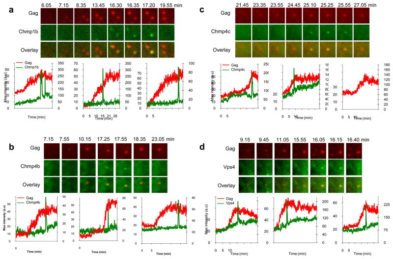
Figure 5. Imaging Chmp1b, Chmp4b, Chmp4c and Vps4A recruitment during EIAV Gag assembly.
Hela cells stably expressing Chmp1b-GFP (a), GFP-Chmp4b (b), GFP-Chmp4c (c) or GFP-Vps4A (d) were transfected with EIAV Gag/Gag-mCherry and observed under TIR-FM beginning at 6 hours post-transfection. Each set of images illustrates the recruitment of GFP-labeled ESCRT proteins during the genesis of an individual VLP. The time after the commencement of observation is given in minutes:seconds. Fields are 2.5x2.5μm. Plots of fluorescence intensity in arbitrary units (a.u) over time for the GFP-ESCRT protein (green, right axis) and Gag-mCherry signals (red, left axis) associated with the assembly of 3 individual VLPs are shown, the left chart in panels a and d correspond to the microscopic images shown above.

