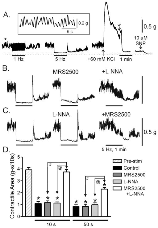
Figure 2. Effects of MRS2500 and L-NNA on nerve induced relaxations in the WT mouse IAS.
(A) Sample traces showing spontaneous phasic and tonic contractile activity along with several experimental procedures. Phasic contractions (*left panel) are shown at a more rapid sweep speed in the inset. 1 Hz (left trace) and 5 Hz (middle trace) EFS gave rise to frequency dependent relaxation. Peak contraction was determined at the end of the experiment with 60 mM KCl (w, wash; right panel) and zero tone with SNP (10 μM) plus nifedipine (1 μM, no further relaxation occurred in this muscle). (B) MRS2500 (1 μM) alone did not reduce relaxation at 5 Hz EFS but rebound contraction was reduced. MRS2500 also did not reduce relaxation with 1 Hz EFS (data not shown). (C) L-NNA (100 μM) alone also did not reduce relaxation but in this case rebound contraction increased. MRS2500 and L-NNA together abolished relaxation for approximately 20–30s of EFS at which point a slow onset relaxation began (B and C). (D) Summary graph of the effects of MRS2500 (n=8) and L-NNA (n=4) on contractile area during EFS. Contractile area during EFS (5 Hz, 60s) is compared to the area preceding EFS (“Pre-stim”, n=12). Contractile area during the first 10s of EFS and the subsequent 50s of EFS are shown separately (all areas have been normalized to area/10s). EFS gave rise to significant (*) relaxation under control conditions and in the presence of either MRS2500 or L-NNA. Neither MRS2500 nor L-NNA alone significantly reduced relaxation during either the initial 10s or the subsequent 50s of EFS, whereas combined MRS2500 and L-NNA (n=8) abolished the EFS-induced relaxation during the initial 10s of EFS and significantly reduced relaxation during the subsequent 50s of EFS (# and @). Shown are mean values ±S.E.M.

