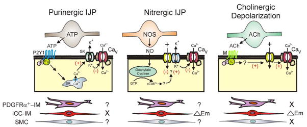
Figure 7. Diagram depicting proposed pathways giving rise to changes in membrane potential with neuromuscular transmission in the mouse IAS.
Shown are inhibitory motor neurons releasing either purines (e.g., ATP) or nitric oxide (NO) and excitatory motor neurons releasing acetylcholine (ACh). The postjunctional cells giving rise to changes in Em are depicted in the center along with proposed second messenger pathways. The three possible cell types considered for these responses are shown at the bottom of the diagram. Left panels: Purinergic IJPs persisted in the W/Wv mouse IAS suggesting that they were independent (X) of ICC-IM leaving either PDGFRα+-IM or SMC as possible candidates (?). ATP or a related purine binds to P2Y1 receptors leading to the release of Ca2+ from the sarcoplasmic reticulum (SR) which activates SK channels (see 14, 36) hyperpolarizing Em and closing L-type Ca2+ channels (Cav). Middle panels: Nitrergic IJPs were greatly diminished in the W/Wv mouse suggesting that ICC-IM play an important role in the generation of these events (△Em). Since nitrergic NMT still blocks slow waves in the W/Wv mouse IAS, other ICC-IM independent pathways are likely involved (?). NO activates guanylate cyclase leading to generation of cGMP. This in turn hyperpolarizes Em via an unknown mechanism that may involve either inhibition of inward current or activation of potassium (K+) channels. Hyperpolarization in turn closes Cav. Right panels: Cholinergic depolarization was only observed in WT mice suggesting that ICC-IM play a critical role in generating this event (△Em) whereas PDGFRα+-IM and SMC do not (X). ACh binds to muscarinic receptors (M) leading to depolarization via an unknown mechanism that perhaps involves stimulation of inward currents. Depolarization in turn opens Cav.

