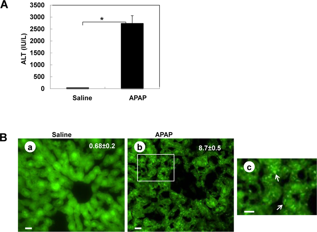
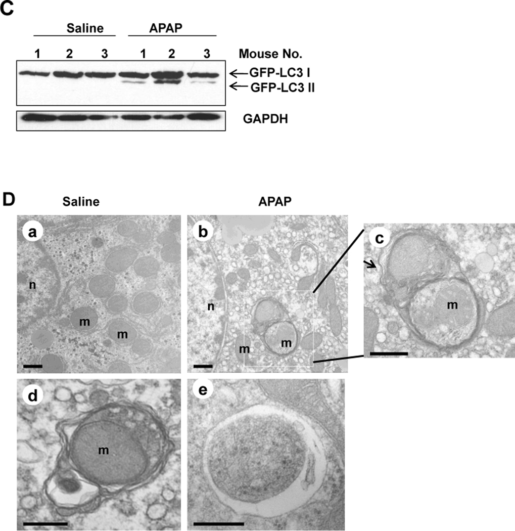
Figure 1. APAP overdose induces autophagy in the liver.
(A–D) GFP-LC3 mice (n=3) were treated either with saline or APAP (500 mg/kg) for 6 hrs, and blood was analyzed for ALT level (A) and the liver sections were analyzed by fluorescence microscopy (B). *: p<0.01. Panel a: saline; panel b: APAP; Panel c is enlarged photograph from the boxed area in panel b. Arrows denote GFP-LC3 puncta. The number of GFP-LC3 puncta (mean ± SEM) was quantified from each animal. More than 30 cells were counted in each individual experiment. Total lysates of the liver were analyzed by immunoblot assay using an anti-GFP antibody (C). (D) Wild-type mice were treated as in (A) and liver samples were processed for EM. Panel a: saline; panel b: APAP, panel c was from the boxed area in panel b; panels d–e: representative EM images of autophagosomes containing mitochondria from APAP treated mouse liver. Arrows denote autophagosomes. n: nucleus; m: mitochondria.


