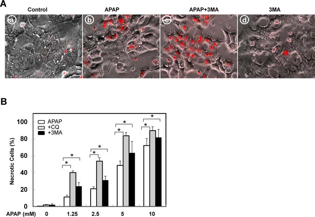
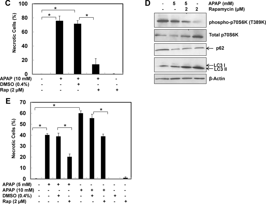
Figure 6. Induction of autophagy protects against APAP-induced cell death.
(A) Representative overlayed images of phase-contrast with PI staining of primary cultured mouse hepatocytes treated for 24 hrs as indicated: panel a: non-treated; panel b: APAP (5 mM); panel c: APAP (5 mM) + 3-MA (10 mM); and panel d: 3-MA (10 mM). (B) Hepatocytes were either treated with saline or with various concentrations of APAP (1.25, 2.5, 5 &10 mM) in the absence or presence of CQ (20 µM) or 3MA (10 mM) for 24 hrs. PI positive cells with intact nuclei were quantified for each experiment (n=3). More than 300 cells were counted in each individual experiment *: p<0.01. (C) Primary mouse hepatocytes were co-treated for 24 hrs as indicated: panel a: DMSO; panel b: APAP (10 mM)+ DMSO (4 µL/mL); panel c: APAP (10 mM) + Rap (2 µM); and panel d: Rap (2 µM). PI positive cells with intact nuclei were quantified for each experiment (n=3). More than 300 cells were counted in each individual experiment *: p<0.01. (D) Primary hepatocytes were treated as indicated for 6 hrs and total cell lysates were subjected to immunoblot assay. (E) Primary mouse hepatocytes were treated with saline or APAP (5 & 10 mM) for 3 hrs, the cells were then further treated with or without DMSO or Rap (2 µM) for another 21 hrs. PI positive cells with intact nuclei were quantified for each experiment (n=3). *: p<0.01.


