
Figure 1. Examples of T cell infiltration in colorectal tumors.
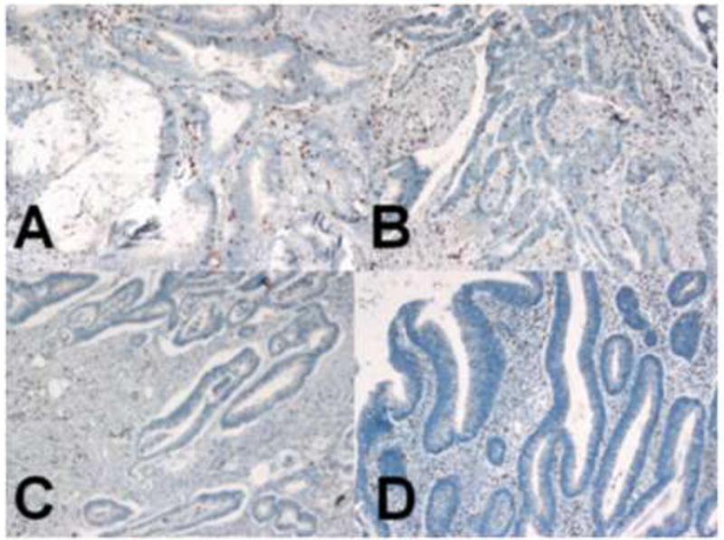
Dark brown colored nuclei indicate CD8+ T cells by anti-CD8 immunohistochemical staining (100X). (A) Poorly-differentiated adenocarcinoma and (B) moderately-differentiated adenocarcinoma showed more prominent CD9+ T cell infiltration in both tumor cell nest and tumor stroma than (C) well-differentiated adenocarcinoma and (D) adenomas.