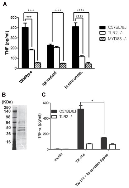
Figure 6.
Induction of TNF-α secretion from macrophages by heat-killed B. anthracis and lipoprotein extracts. (A) Bone marrow-derived macrophages from the three types of mice indicated were exposed to heat-killed wild-type strain, lgt mutant, or complemented mutant at 100 μg ml−1. Macrophages were incubated for 18 h and supernatants were analyzed for TNF-α by enzyme-linked immunosorbent assay. Data are plotted as the mean values ± standard deviation of three independent experiments. Significant differences where p < 0.001 are indicated by three asterisks (***) and where p < 0.0001, indicated by four asterisks (****). (B) Coomassie blue staining of proteins in TX-114 fraction of B. anthracis (second lane). First lane contains molecular weight markers. (C) C57BL/6J bone marrow-derived macrophages from the two types of mice indicated were incubated with medium, TX-114 fraction, or lipoprotein lipase digested TX114 fraction for 18 h and supernatants were analyzed for TNF-α by enzyme-linked immunosorbent assay. Data are plotted as the mean values ± standard deviation of three independent experiments. Significant difference (p < 0.05) between lipoprotein lipase treatment and no treatment is indicated by an asterisk (*). All statistical tests used the unpaired two-tailed t-test.

