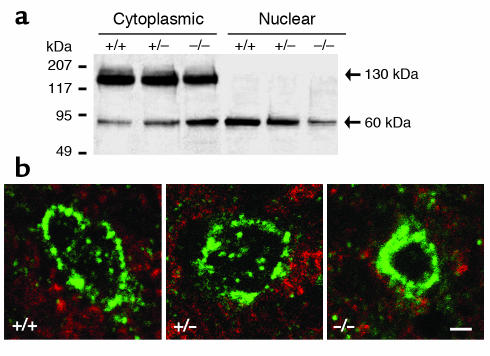
Figure 7.
SREBP1 localization in LV cardiomyocytes of mice aged 4–6 weeks. (a) Western blot analysis of cytoplasmic and nuclear fractions of LV tissue hybridized with an SREBP1 Ab. Representative data for one WT (+/+), Lmna+/– (+/–), and Lmna–/– (–/–) mouse are shown. Experiments were repeated with six mice from each group. When compared with WT cardiomyocytes, Lmna–/– cardiomyocytes show a mean decrease (35%) in uncleaved (130 kDa) SREBP1 and a mean increase (55%) in cleaved (60 kDa) SREBP1 in the cytoplasmic fraction, with a mean reduction (40%) of cleaved SREBP1 in the nuclear fraction. (b) Immunofluorescence microscopy shows a perinuclear and intranuclear distribution of SREBP1 in WT and Lmna+/– nuclei, but predominant perinuclear SREBP1 staining in Lmna–/– nuclei. Scale bar: 2 μm.

