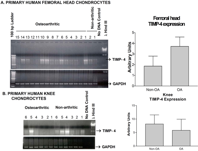
Fig. (2).
A) Expression of the TIMP-4 mRNA in primary human non-arthritic and OA femoral head articular chondrocytes. All conditions were same as in Figure 1. The right panel is a cumulative graphic depiction of mean ± SEM TIMP-4/GAPDH ratios indicating TIMP-4 increase in hip OA chondrocytes. B) TIMP-4 RNA expression by RT-PCR analysis in knee chondrocytes from six non-OA and 6 OA patients with negative control (no DNA) and markers. The right panel is a cumulative graphic depiction of mean ± SEM TIMP-4/GAPDH ratios indicating TIMP-4 decrease in knee OA chondrocytes.

