Figure 2.
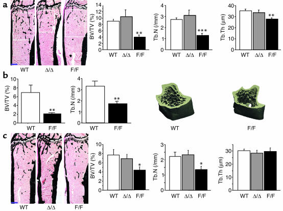
Low trabecular bone volume in male and female gp130Y757F/Y757F mutant mice. (a) Representative images of von Kossa-stained tibiae of male 16-week-old WT, Δ/Δ, and F/F mutant mice, showing differences in trabecular bone volume (white boxes show secondary spongiosa region used for histomorphometric measurements; note that this is a smaller anatomical region in the Δ/Δ mice due to the smaller bone size). Scale bar (blue), 500 μm. Histomorphometric measures of BV/TV, Tb.N, and Tb.Th were all reduced in F/F mice compared with WT and Δ/Δ mice. (b) Micro-CT analysis and representative three-dimensional images of tibiae from male WT and F/F mice. (c) Representative images and histomorphometry for female 16-week-old WT and gp130 mutant mice. Scale bar (blue), 500 μm. BV/TV is reduced in female F/F mice compared with WT and Δ/Δ mice. All values are mean ± SEM from a minimum of seven mice per group at 12–16 weeks of age for histomorphometry and 8–12 weeks of age for micro-CT. *P < 0.05; **P < 0.01; ***P < 0.001 vs. WT of the same sex.

