Figure 4.
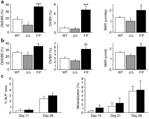
Increased bone formation in male and female gp130Y757F/Y757F mutant mice. Histomorphometric indices of bone formation including tibial ObS/BS, OV/BV, and MAR were significantly higher in male (a) and female (b) F/F mice compared with WT and Δ/Δ mice. All values are mean ± SEM from a minimum of eight mice per group at 12–16 weeks of age. *P < 0.05; **P < 0.01; ***P < 0.001 vs. WT of the same sex. (c) Ex vivo osteoblast differentiation from bone marrow cultured under osteoblastogenic conditions was not significantly altered in F/F mice (black bars) compared with WT mice (white bars). Shown are alkaline phosphatase–positive colony formation as a percentage of well area (%ALP+ area) and mineralization detected by a von Kossa stain for calcified matrix (%). Values are mean ± SEM from three experimental preparations at each time point using a minimum of three mice of mixed sexes per genotype for each experiment.

