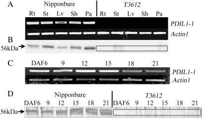
Fig. 5.
Expression of PDIL1-1. (A) Expression as assayed by RT-PCR. Actin1 was used as the reference. Rt, root; St, stem; Lv, leaf; Sh, sheath; Pa, panicle. (B) Western blot analysis based on anti-PDIL1-1 polyclonal antibodies. The absence of PDIL1-1 protein in the mutant is highlighted. (C) Expression of PDIL1-1 in the developing [6–21 days after flowering (DAF)] endosperm of cv. Nipponbare assayed by RT-PCR. (D) Western blot analysis of the presence of PDIL1-1 in the developing endosperm. The absence of PDIL1-1 protein in the mutant is highlighted.

