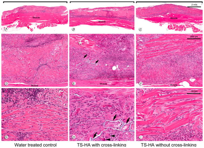
Fig. 2.
Representative H&E images at 1 month of: a, d, g water treated control; b, e, h TS-HA with cross-linking; and c, f, i TS-HA without cross-linking. a–c (×10) Fascia grafts (brackets) were still visible and discernable from the underlying abdominal skeletal muscle. d–f (×100) Remnant fascia architecture was identifiable as longitudinal, collagenous bands. Grafts exhibited a variable distribution of cellular infiltrates and acellular, collagenous regions (asterisk), as demonstrated in treated fascia with cross-linking (e). g–i (×400) Grafts were heavily infiltrated by chronic inflammatory cells, which were predominantly lymphocytes, as demonstrated in water controls (g). In cross-linked treated fascia, TS-HA hydrogel was discernable (arrows) and surrounded by multinucleated giant cells (h, arrowheads). Spindle-shaped, fibroblast-like cells heavily infiltrated the grafts, often between the collagenous bands, as demonstrated in uncross-linked treated fascia (i)

