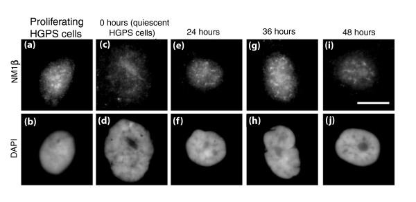
Figure 6.
Myosin staining pattern in quiescent HGPS human dermal fibroblasts following re-stimulation. HDFs from HGPS patient AG11498 were serum-starved for 7 days to induce quiescence. The cells were then re-stimulated with fresh serum and samples were collected at 0, 24, 36 and 48 hours post-serum restoration. Samples were also collected before serum withdrawal (proliferating cells). The samples were then fixed with methanol:acetone (1:1) and distribution of NM1β was assessed by performing a dual color indirect immunofluorescence assay for NM1β (a, c, e, g, i) and pKi67 (b, d, f, h, j). (a, c, e, g, i) The distribution of NM1β in cells before and after re-stimulation of quiescent fibroblasts. Scale bar = 10 μm.

