
Figure 4.
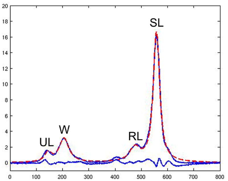
1H-MRS of the vertebral bone marrow resolves four peaks: unsaturated lipids (UL), water (W), residual lipids (RL) and saturated lipids (SL). The four peaks were fitted with Voigt models to estimate their areas and line widths (18;19). The acquired spectrum (blue line with the four peaks) is overlaid with the fitted spectrum (dashed red line). The lower blue line indicates the difference between acquired and fitted spectrum.