Figure 1.
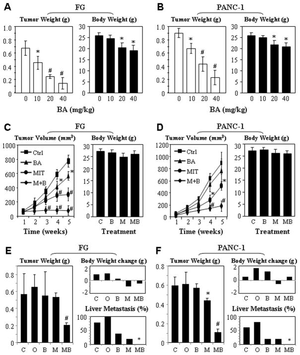
Dose-dependent antitumor effects of BA and MIT in xenograft models of human pancreatic cancer. Dose response: FG (A) and PANC-1 (B) cells were injected into the pancreases of nude mice (n=5). Ten days after tumor injections, the mice were treated with different doses of BA (10, 20, and 40 mg/kg) via oral gavages three times a week. The tumors were weighed 45 days after tumor cell injection (left panels); the mice were weighed at the same time and columns, mean weights; bars, standard deviations (right panels). Synergistic antitumor effect in ectopic models: FG (C) and PANC-1 (D) cells were injected into the subcutis of nude mice (n=5). When tumors reached around 4 mm in diameter, the animals received MIT (0.05 mg/kg) via intraperitoneal injection twice a week and BA (10 mg/kg). Tumor volumes were measured every week until the mice were killed 45 days after tumor cell injection (left panels); the mice were weighed at the time of termination of experiments (right panels). Synergistic antitumor effect in orthotopic models: FG (E) and PANC-1 (F) cells were injected into the pancreases of nude mice (n=5). Ten days after tumor cell injections, the animals received MIT (0.05 mg/kg) via intraperitoneal injection twice a week and BA (10 mg/kg) via oral gavage three times a week. The mice were killed 45 days after tumor cell injection; both tumors (left panels) and the mice (upper right panels) were weighed, and hepatic metastases were determined (lower right panels). *P<0.05 and #P<0.01 as compared to respective controls (two tailed student t test). C, Control; O, Corn oil; B, BA; M, MIT; MB, MIT+BA.

