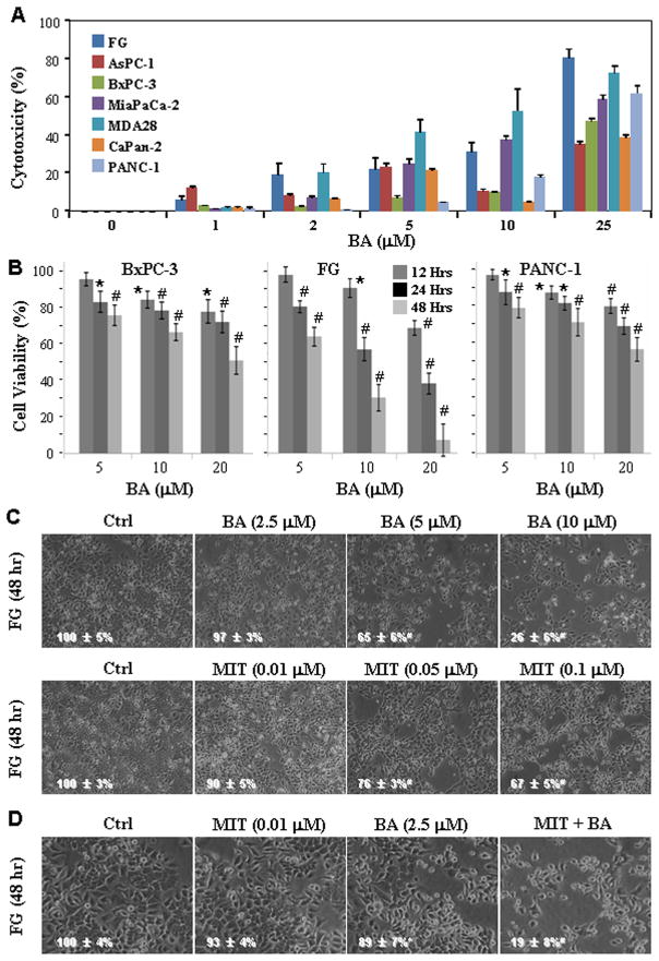
Figure 2.
Synergistic effect of treatment with BA and MIT on inhibition of pancreatic cancer cell proliferation. A, Various pancreatic cancer cell lines were treated with BA at concentrations ranging from 1 to 25μM for 48 h. Inhibition of cell proliferation was assessed using an MTT assay. B, BxPC-3, FG, and PANC-1 cells were treated with BA at concentrations ranging from 5, 10, and 20μM for 12, 24 or 48 h. Inhibition of cell proliferation was assessed using an MTT assay. C, FG cells were treated with BA at concentrations ranging from 2.5, 5, and 10μM or MIT at concentrations ranging from 0.01, 0.05, and 0.10μM for 48 h. Cell cultures were photographed before assessing cell proliferation using an MTT assay (inserted number represented percent viability ± SD). D, FG cells were treated with 2.5μM BA, 0.01 μM MIT or both for 48 h. Cell cultures were photographed before assessing cell proliferation using an MTT assay (inserted number represented percent viability ± SD). *P<0.05 and #P<0.01 (two-tailed student t test).

