Figure 6.
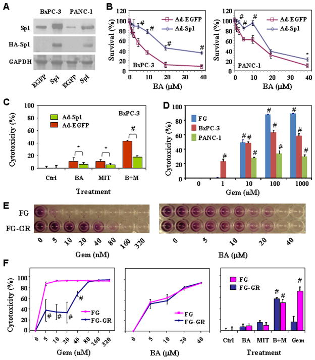
Influence of Sp1 expression on cytotoxicity in vitro. BxPC-3 and PANC-1 cells were treated with Ad-EGFP or Ad-Sp1 (10 MOI) for 6 hours, and cultures were incubated for additional 18 h. The cells were either harvested for analysis of Sp1 expression by using Western blot (A) or plated in 96-well plates and treated with different concentrations of BA (B) or with 2.5 μM BA, 0.01 μM MIT or both (C) for additional 24 h before cell viability determination by MTT assay. D, BxPC-3, FG, and PANC-1 cells were treated with gemcitabine (“Gem”, 0 – 1000 nM) for 48 h. Inhibition of cell proliferation was assessed using an MTT assay. E & F, FG and FG-GR (gemcitabine-resistant variant) cells were treated with gemcitabine (0 – 320 nM) or BA (0 – 40 μM) for 72 h or treated with 2.5 μM BA, 0.01 μM MIT or both for 48 h. Treatment with 10 nM of gemcitabine was used as a control. Inhibition of cell proliferation was assessed using an MTT assay. *P<0.05 and #P<0.01 as compared to respective controls (two tailed student t test).

