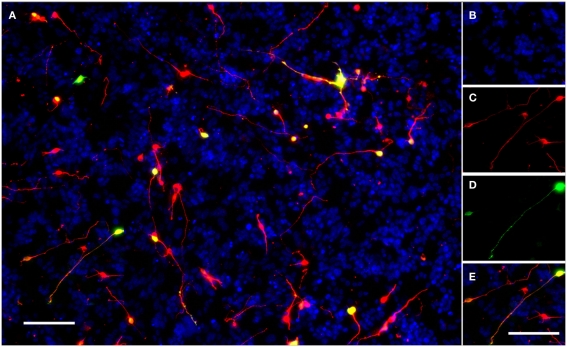
Figure 1.
A typical field of view showing electroporated CGNs cultured on a monolayer of growth-permissive CHO-R2 cells. (A) A typical field of view taken using the IN Cell Analyzer 1000 semi-automated cell imager with a 10× Nikon ApoPlan objective. (B) DAPI stained nuclei of CHO-R2 cells and CGNs. (C) CGNs stained for beta-III-tubulin. (D) eGFP positive, transfected CGNs expressing GFP. (E) The merged image; transfected neurons appear yellow. Scale bars: 100 μm.

