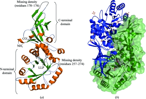
Figure 1.
(a) Structure of the S. aureus alanine racemase monomer. Ribbon representation with α-helices coloured orange and β-sheets shown in green. The PLP cofactor covalently bound to Lys39 is shown as a black stick model. (b) Ribbon representation of the S. aureus alanine racemase dimer. Monomers are coloured blue and green, with the surface representation of one monomer also shown in green. The PLP cofactors are depicted as black stick models. Sulfate and acetate molecules are shown as ball-and-stick models; C atoms are coloured black, O atoms red, S atoms yellow and phosphates orange.

