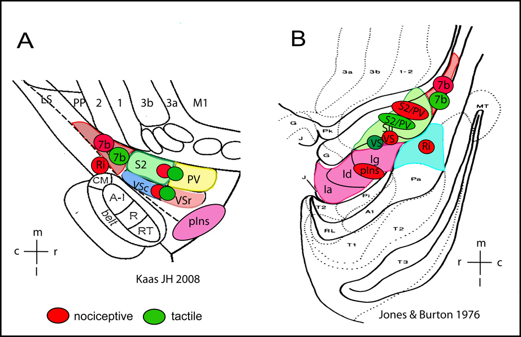
Figure 6.
Schematic summary of cortical areas that are responsive to nociceptive heat (red patches) and tactile (green patches) stimuli in anesthetized squirrel monkeys. Responsive cortical areas are indicated by color circles and placed over the schematic illustrations of cortical areas near the lateral sulcus in New World monkeys as established by two research groups (adapted from Kaas JH 2008 (A) and Jones & Burton 1976(B)). A: electrophysiologically and cytoarchitectonically defined cortical areas in New World monkeys. Ri: retroinsula; insula; Ins: insula; S2: secondary somatosensory area; PV: parietal ventral area; VSr: rostral ventral somatosensory area; VSc: caudal ventral somatosensory area; 7b: area 7b. m: medial; l: lateral; c: caudal; r: rostral. B: Cytoarchitectonically defined cortical areas in squirrel monkeys. SII includes subregions of S2, PV, VSc, VSr in Kaas 2008 (A). Insular cortex is divided into three subregions: Ig (granular field), Id (dysgranular field), and Ia (agranular field). Ri is located posterior to Insular region.
High-resolution fMRI revealed distinct and shared nociceptive heat and innocuous tactile processing networks within posterior parasylvian region of monkeys

