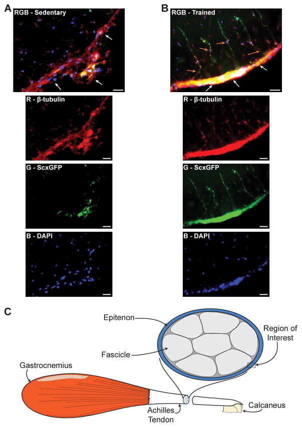
Figure 5.
Compared with sedentary mice (A), mice that underwent treadmill training (B) displayed a dramatic increase in scleraxis expression in the epitenon (white arrows) and an emergence of fibroblasts from the epitenon (orange arrows). Composite images (RGB) are in the top panel, and the individual red, green and blue channels are below. There was substantial overlap between β-tubulin and scleraxis signal in the epitenon (yellow). An illustration demonstrating the region of interest is shown in (C). β-tubulin, red; Scleraxis-GFP, green; nuclei, blue. Scale bars = 20μm.

