Fig. 2.
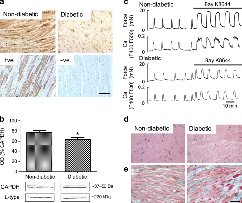
Uterine histology and Ca channels. a Tissue distribution of L-type calcium channels. Immunohistochemical localisation of L-type calcium channels in non-diabetic and diabetic myometrium with positive and negative controls underneath. Scale bar, 50 μm. b Quantification of L-type calcium channel expression. Densitometric western blot quantification of L-type calcium channels in non-diabetic and diabetic patients. c Effect of Bay K-8644 stimulation. Simultaneous measurements of force and Ca (Indo-1) in myometrium from non-diabetic and diabetic patients during spontaneous activity and with application of 1 μmol/l of the Ca channel agonist, Bay K-8644. d and e Myometrial histology. d H&E. e Masson’s Trichrome-stained uterine sections from non-diabetic and diabetic patients. Scale bar, 50 μm. Significant difference, *p < 0.05

