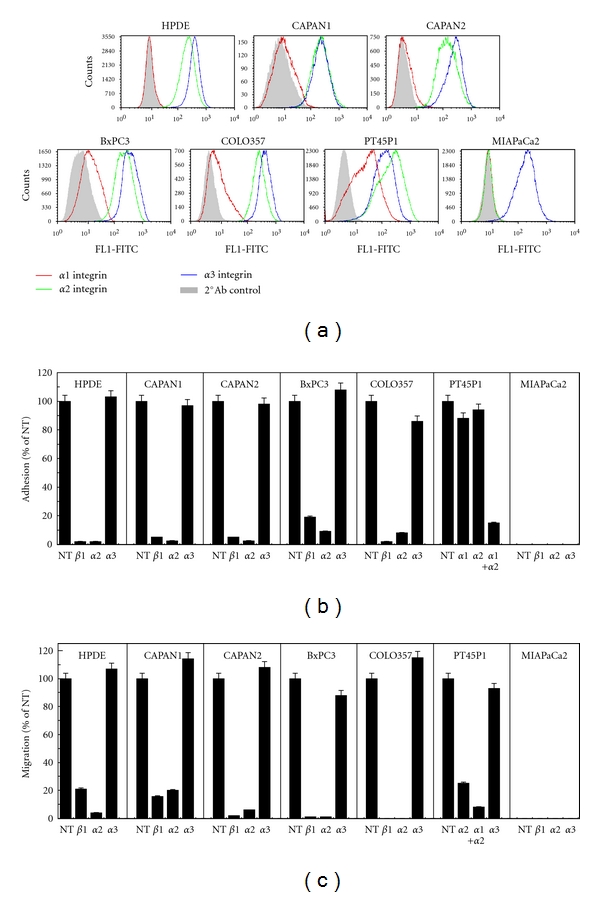
Figure 1.

PDAC integrin expression and utilization reflects cellular differentiation. (a) Flow cytometric analysis of α1, α2, or α3 surface expression in a spectrum of pancreatic ductal cells including untransformed (HPDE), well-(CAPAN1 and CAPAN2), moderately (BxPC3, COLO357) and poorly differentiated PDAC (PT45P1 and MIAPaCa2) (see Supplemental Table S1 for cell characteristics). Secondary antibody controls, solid. (b) Adhesion of the cells in (a) to 25 μg/mL collagenI for 45 minutes in the presence or absence of function-blocking antibodies to the indicated integrins, as described in Materials and Methods. (c) Serum-free migration (16 h) through Transwell inserts coated on the underside with 20 μg/mL collagenI in the presence or absence of function-blocking antibodies as in (b).
