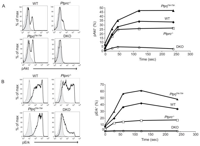
Figure 5. CD45 and CD148 differentially regulate signaling events following fMLF stimulation.
(A) BM cells from mice of the indicated genotypes were stimulated with fMLF (0.2 μM) for indicated time, fixed, permeabilized, and then stained with phospho-Akt along with other surface makers. Neutrophils were identified as CD11b+Gr1+. Phospho-Akt of stimulated (2.5 min, shown as black lines) and control samples (shown as filled histograms) were overlaid. Percentages of pAkt+ cells over a time course are shown on the right side. Data are representative of three independent experiments. (B) BM cells were treated same as in (A) and phospho-ERK was stained similarly. Percentages of pERK+ cells over a time course are shown on the right side. Data are representative of three independent experiments.

