Figure 1.
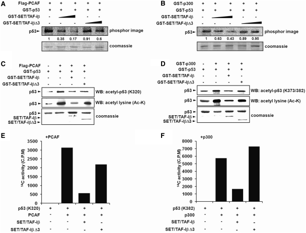
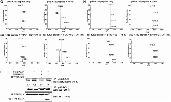
SET/TAF-Iβ inhibits PCAF- and p300-mediated p53 acetylation. (A and B) Acetylation assays of p53 with PCAF and p300 were performed with increasing concentrations of SET/TAF-Iβ or SET/TAF-Iβ-Δ3. Autoradiogram of INHAT assay using recombinant PCAF (A) or p300 (B) with SET/TAF-Iβ and SET/TAF-IβΔ3 on GST-p53 (top) followed by coomassie staining (bottom). Numbers below phosphorimage represent quantification of p53 acetylation. (C and D) Western blot analysis of in vitro p53-acetylation assay with PCAF (C) or p300 (D), GST-p53, GST-SET/TAF-Iβ or GST-SET/TAF-IβΔ3 using anti-acetyl-p53 (K320) or anti-acetyl-p53 (K373/382) and anti-acetyl lysine. The levels of GST-SET/TAF-Iβ and SET/TAF-Iβ-Δ3 are shown in the bottom panel by coomassie staining. (E and F) p53 peptides (p53-K320 and p53-K382) were used as substrates in the INHAT assay with PCAF (E) or p300 (F) with GST-SET/TAF-Iβ or SET/TAF-Iβ-Δ3. Acetylation levels were quantified via filter binding assay and represented as raw counts per minute (cpm) incorporated. (G and H) p53 peptides were incubated with PCAF (G) or p300 (H) with GST-SET/TAF-Iβ, and the modified peptides were analyzed via LC–MS spectrometry. (I) HCT116 cells were transfected with various plasmids, as indicated, and p53 immunoprecipitates were subjected to western blotting using anti-acetyl lysine. Quantification of acetylated p53 band intensities is shown below. The amounts of immunoprecipitated p53 and transfected SET/TAF-Iβ proteins were determined by western blotting and shown in the lower panels.


