
Figure 2. Feeding increases the size and abundance of individual stem cell clones.
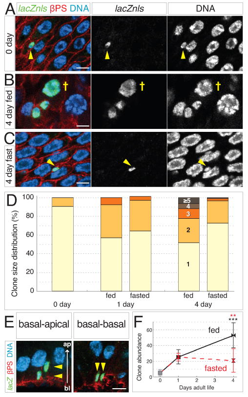
(A-C) Stem cell clones grow larger in fed guts. Clones labeled with tub-lacZnls in green; DNA is blue and βPS integrin outlines cells in red. (A) At 0 days, most labeled cells are single, stem-like cells (arrowhead). (B) In 4-day fed guts, multicell clones are numerous and include polyploid enterocytes (cross). (C) In 4-day fasted guts, most labeled cells remain as single stem-like cells (arrowhead). Scale bars, 5 μm.
(D) Feeding promotes clone growth. At 0 day, nearly all clones contain one cell. In 1-day fed and fasted guts, the proportion of 2-cell clones increases. In 4-day fed guts, clones containing 3 or more cells have become more prevalent. In 4-day fasted guts, 1-cell clones remain predominant. See also Supplemental Table S2.
(E) In cross sections of the gut epithelium, 2-cell clones exhibit either basal-apical (left) or basal-basal arrangements (right). Scale bar, 5 μm.
(F) Feeding increases clone abundance. Between 0 and 1 day, clone abundance (number of discrete clones per distal hairpin) rises 5-fold in fed and fasted guts. In 4-day fed guts, clone abundance has increased to 10-fold higher than at 0 day. In 4-day fasted guts, no further increase occurs. Data are means ± S.D. and obtained from same guts as (D). Black asterisks, p=0.0004 (0 day and 4 day fed); red asterisks, p=0.0026 (4 day fasted and 4 day fed). See also Supplemental Table S2.