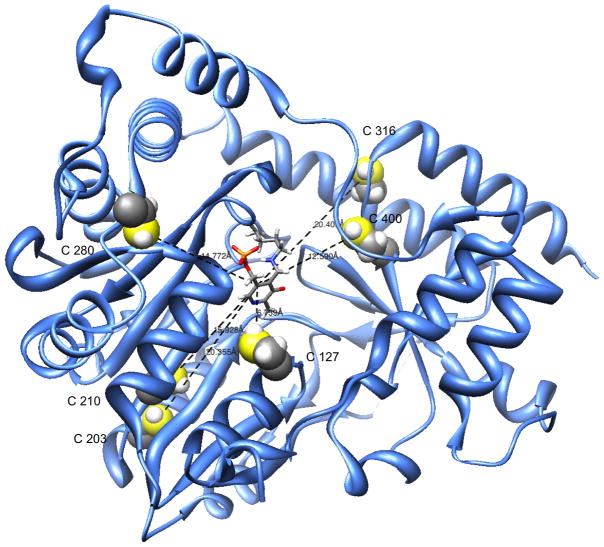
Fig. 4.
Ribbon model of rhGTK monomer showing pyridoxal 5′-phosphate in the active site. The diagram shows a relatively large active site. Cysteine residues are emphasized. Atoms: S, yellow; H, light gray. The model was produced using the UCSF Chimera program ( http://www.cgl.ucsf.edu/chimera/).

