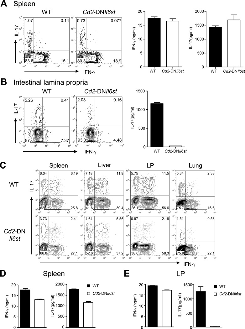
Figure 3. Differential requirement of IL-6 for Th17 cell lineage commitment in different priming micro-environments under lymphopenic conditions.
(A) Splenic naïve (CD62LhiCD44lo) and memory (CD62LloCD44hi) CD4+ T cells from WT and Cd2-DNIl6st Tg mice were sorted and stimulated with PMA and ionomycin stained for intracellular IFN-γ and IL-17 (left two panels). Naïve and memory cells were also stimulated with plate bound anti-CD3 and anti-CD28 for 48 hours and culture supernatants were assayed for IFN-γ and IL-17 (Right two panels). Data for memory cells are shown. Naïve cells did not secrete detectable IFN-γ or IL-17. (B) LPLs were isolated from WT and Cd2-DNIl6st Tg mice and were either activated with PMA and ionomycin followed by staining for intracellular IL-17 and IFN-γ (left panels) or cultured in the presence of plate-coated anti-CD3 and anti-CD28 for 48 hours after enrichment for CD4+ T cells and assayed for IL-17 secretion in the culture supernatants (right panel). (C–E) Cells from the spleen and lymph nodes of WT or Cd2-DNIl6st Tg mice were transferred intravenously into Rag1−/− recipients. After 7 days, mononuclear cells (MCs) were isolated from the spleen, liver, LP and lung. (C) MCs from the indicated organs were stimulated with PMA and ionomycin and stained for intracellular IFN-γ and IL-17. CD4+TCRβ+ positive cells expressing intracellular IFN-γ and IL-17 are shown. (D-E) CD4+ T cells from indicated organs were stimulated with plate-bound anti-CD3 and anti-CD28 for 48 hours and analyzed for IFN-γ and IL-17 secretion in the culture supernatants. Data are representative of two or three independent experiments. Bar graphs represent mean ± SEM.

