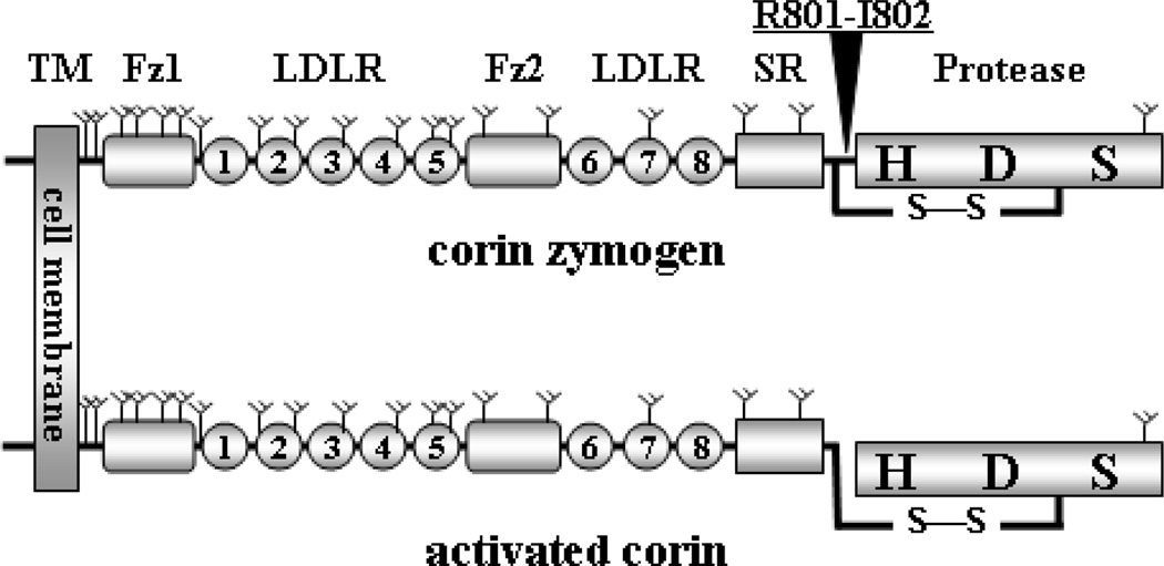
Fig. 1.
Corin protein domain structure. The transmembrane domain (TM), frizzled-like domains (Fz), LDLR repeats, scavenger receptor-like domain (SR), and protease domain (Protease) with active site residues histidine (H), aspartate (D), and serine (S) are indicated. Y-shaped symbols indicate predicted N-glycosylation sites. An arrow head indicates the activation cleavage site between Arg801-Ile802. A disulfide bond (S-S) connects the protease domain and the rest of the molecule after corin zymogen (upper) is activated (lower).

