Figure 3.
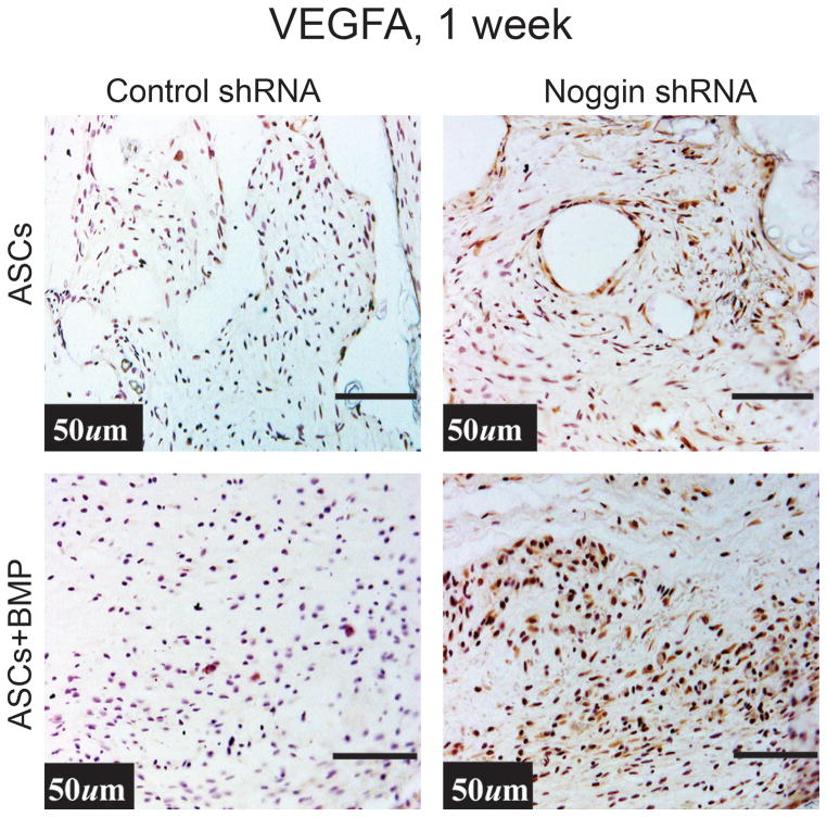
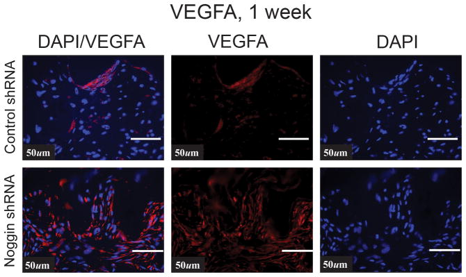
(A) Human VEGF immunohistochemistry in a calvarial defect (brown color) with control shRNA or Noggin shRNA hASCs on a HA coated PLGA scaffold with or without BMP-2 (200ng/ml). (B) VEGFA expression by immunofluorsence (red color) in the region of the calvarial defect treated either with control shRNA treated or Noggin shRNA treated hASCs.


